01 June 2007

Mad scientists cure mad cows: gene-ious

Mad cow disease(MCD) is a neurodegenerative disease that affects bovines. If infected meat is consumed it can be passed onto humans in the form of Cruetzfeldt-Jakob Disease, which also affects the brain. It is fatal to both.
MCD is caused by the misfolding of a normal cellular protein (prpc), into an infectious protein, a prion.
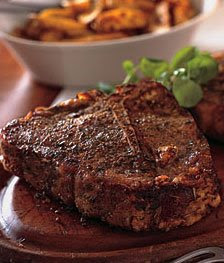
Due to the fact that everyone (almost) loves a nice steak, MCD in cows is the main problem. Richt et al have used genetics to disrupt the alleles of prpc, producing 12 cloned calves that are prpc deficient. The cows were proved to be immune to MCD after prions were injected into the brain tissue of two of the cows with no effect. The remaining cows were injected with MCD with no effect aswell, but as MCD can lie dormant for several years so, time will tell if they are truly immune.
‘At over 20 months of age they [the cows] are clinically, physiologically, histopathologically, immunologically and reproductively normal’ (Richt et al, 2007).
Aside from reducing/eliminating the occurrence of MCD, these cattle are thought to be beneficial for future drug and prion research. They may also produce favourable, prion free products such as the steak mentioned earlier.
So, no mad cows, no mad people, what’s the problem? Certain issues have been raised by the experiment. Will human consumption of the cows cause side effects? Will reproduction with normal cows cause problems? Is it moral to ‘play god’ by changing and controlling another species mainly for our own benefit?

References:
Primary:
Richt JA, Kasinathan P, Hamir AN, Castilla J, Sathiyaseelan T, Vargas F, Sathiyaseelan J, Wu H, Matsushita H, Koster J, Kato S, Ishida I, Soto C, Robl JM & Kuroiwa Y, 2007, Production of cattle lacking prion protein, Nature Biotechnolgy, vol 25, pp 132-138
Secondary:
Paddock C, 2007, ‘Could Genetic Engineering Eradicate Mad Cow Disease?’, 27th May 2007
Canadian Food Inspection Agency, 2007, ‘BSE Case Confirmed in British Columbia’, 30th May 2007

Not far from a triumphant victory over mastitis in dairy cows


Mastitis in dairy cows causes huge economic loss in the global dairy industry. Despite various measures has been adopted to tackle mastitis, its occurrence has not been declined significantly. A recent research developed a possible solution by creating transgenic cows with the enzyme lysostaphin secreted in their milk. Transgenic cows carrying lysostaphin in the study has shown to be more resistant to S.aureus, a major contagious mastitis pathogen which accounts for 30% of all intramammary infections. While lysostaphin effectively eliminates the pathogen S.aureus, it is relatively harmless to both the cows and the milk consumers compared with commonly used antibiotics. Furthermore, it has minimal effect on milk composition.

To maximize the efficiency in tackling mastitis in dairy cows, major pathogens other than S.aureus need to be dealt with also. Antimicrobials which satisfy a series of criteria, including harmlessness to cows and milk consumers, no biological activity when taken orally and high sensitivity to pathogens, are hard to find. Nevertheless, the development of transgenic cows that secrete antimicrobials in milk is a breakthrough in the field of dairy science and surely of great benefit to the global dairy industry.

Aiming at an increase in milk yield and improvement in milk quality, more dairy science research in creating mastitis-resistant cows are expected to be done in the near future.

Primary source:
Donovan, D.M., Kerr, D.E., Wall, R.J., 2005. ‘Engineering disease resistant cattle’, Department of Animal Science, University of Vermont, Burlington
http://www.springerlink.com/content/u1m28j7732815g68/fulltext.pdf

Secondary source:
Kerr, D.E., Wellnitz, O., 2003. ‘Mammary expression of new genes to combat mastitis’, Department of Animal Science, University of Vermont, Burlington
http://jas.fass.org/cgi/reprint/81/suppl_3/38.pdf

Written by: Kin Fai, Lau
Student number: 41281941

Future of champion stallions

Future of champion stallions

In April 2005 at a press conference in Italy the first ever clone of a champion racehorse was unveiled. Italian scientists cloned Pieraz who had won two world endurance titles. Pieraz had been castrated at a young age like most successful endurance horses. The aim of this clone was to preserve Pieraz genetic heritage.
As Professor Galli leader of the project said “What we have done is to create a clone of a gelding that is a stallion, to preserve the genetic heritage of an exceptional champion. This gives us an unprecedented opportunity to breed from the best animals.” To clone Pieraz a sample of cells from the champion was taken in 2002 and stored in liquid nitrogen after an agreement between Ms Kanavy the owner and Cryozootech, a biotechnology company based near Paris. Scientists from Cryozootech and CIZ a breeding animal company then cloned Pieraz. The horse was cloned using the cell nuclear transfer technique which was developed at the Roslin Institute, near Edinburgh. This is the technique which led to the birth of Dolly the cloned sheep. Although Pieraz-Cryozootech-Stallion the cloned foal was born perfectly healthy there is no guarantee that he will perform as well as Pieraz. If this clone appears to be able to breed it can lead to a new generation of cloned champion stallion.

References

Henderson, M. 2005, Champion the wonder clone, Times online
http://www.timesonline.co.uk/tol/news/world/article381252.ece (accessed 28/05/07)

Coghlan, A. 2005, First clone of champion racehorse revealed, New scientist.
http://www.newscientist.com/article.ns?id=dn7265 (accessed 28/05/07)

When Neutral Bugs Turn Nasty: Venezuelan equine encephalitis virus


The Venezuelan equine encephalitis virus (VEEV) is responsible for a zoonic disease transmitted by mosquitoes that can cause death in humans and equines (such as horses, donkeys and asses). This disease emerges periodically throughout central and southern America and occasionally even in the USA. In humans it causes flu-like symptoms including severe headaches which usually subside after a few days. Death usually only occurs in immunocompromised people such as the very old or young. In equines the effects are more severe and neurological disorders are often observed. The death rate in equines has been estimated as high as 83% for some strains. In these animals death usually occurs within a week of infection.

It has been hypothesised that VEE epidemics are caused by alterations to the genome of a relatively harmless strain of the virus. This less virulent strain of the virus is carried asymptomatically by many small, forest dwelling mammals throughout the Americas and is only capable of infecting humans who are continuously in close proximity with infected populations. This strain is very rarely passed to equines. It was shown in a recent study that a single mutation of the genome causing an amino acid substitution in the less virulent strain is all that is needed to turn it into an epidemic strain. Because of this, it is believed that these outbreaks of highly virulent VEEV will continue to occur unless mosquito populations are better controlled and more equines are vaccinated.


Written by: Sarah Batt

Student number: 41386015


Primary references:
http://www.pubmedcentral.nih.gov/articlerender.fcgi?tool=pubmed&pubmedid=16549790 Venezuelan encephalitis emergence meditated by a phylogenetically predicted viral mutation

http://en.wikipedia.org/wiki/Venezuelan_equine_encephalitis_virus -general info from wikipedia

Secondary references:
http://arjournals.annualreviews.org/doi/pdf/10.1146/annurev.ento.49.061802.123422 - more in depth info on outbreaks

http://www.cdc.gov/ncidod/EID/vol11no05/04-1251.htm -about reservoirs (forest mammals)

http://www.sciencedaily.com/releases/2006/09/060915203515.htm -bioterrorism threat

The Great Debate – Drumstick or Wing?

Scientists, it seems, have decidedly taken sides with the drumstick lovers and have created a chook with three legs.

They have been working on trying to unravel the genetic sequence of limb development, that is – why is a leg a leg and not a wing? They focused on three genes in particular. Normally, Tbx5 is turned on in the wing, and Tbx4 and Pitx1 are turned on in the leg.

To determine the gene responsible for leg development the scientists stuck Pitx1 into a virus, then sprayed the virus on the would-be wing nub of a chick embryo. Amazingly, the resulting chicken had 3 legs and just one lonely wing!

However, the third leg isn’t a perfect drumstick. While feathers became scales, and the limb was straight, clawed and muscled like a leg, some wing characteristics still remained.

This means that Pitx1, while switching on the ‘leg gene’ (Tbx4) in the wing, had no effect on the actual ‘wing gene’ (Tbx5), being independent of it. Both were switched on, making the limb a wing-leg hybrid. More bizarrely, Japanese researchers have now achieved a complete wing and leg position interchange.

While these odd chooks have caused egg-citement in chook farmers and KFC lovers alike, their creation had a more noble purpose. Scientists believe that by understanding the genes responsible for forelimb and hindlimb development, they could one day be able to correct or prevent limb deformities in humans.

Really though, my vote is for the four winged chook. Less meaty, a little less able to walk, but altogether a major step back towards that airy domain, so that they may fulfill their ancestry and return to their rightful place as lords of the sky.

by Stephanie Clark

41207268

Genetics of a cow gone mad


Mad cow disease, also known as Bovine Spongiform Encephalopathy (BSE) is termed a prion disease and variants of this abnormal protein occur in humans through consumption of contaminated meat as well as other livestock. As the term prion suggests, this disease is a result of polymorphisms in the coding and regulatory regions of the normal prion protein (PRNP) gene localized on chromosome BTA 13 at position q17.

BSE is not a viral or bacterial infection because the alteration in the DNA sequence is brought about by an infectious prion protein acquired primarily from contaminated feed. Furthermore, scientists have proven that only cows which are genetically susceptible are affected. So far, studies have shown that this susceptibility factor is highly associated with the expression of other genes in chromosomes 5, 10 and 20 in addition to the PRNP locus.

In a case study where they compared the genes of healthy and infected cattle, they found that diseased cattle across different breeds were characterized by a higher frequency of insertion/deletion polymorphism within the open reading frame of the PRNP gene.

The body naturally has normal prion proteins within its cells which the infectious prion converts into a form identical to its own after entering. The accumulation of these indigestible proteins leads to chronic degeneration of the central nervous system. Clinical symptoms include ‘nervousness’, kicking, abnormal walking and pelvic limb ataxia.

Despite these advances, scientists are still working to uncover the puzzling mechanism behind the concept of an infectious ‘protein’.


References
Primary source –

Zhang, C., de Koning, D-L., Hernandez-Sanchez, J., Haley, C.S., Williams, J.L. & Wiener, P. 2004, ‘Mapping of Quantitative trait loci affecting Bovine Spongiform Encephalopathy’, Genetics Society of America [online] Available at: http://www.ncbi.nlm.nih.gov/sites/entrez?Db=pubmed&Cmd=ShowDetailView&TermToSearch=15342524 (accessed 28/05/07)

Secondary source –
Czarnik U, Zabolewicz T, Strychalski J, Grzybowski G, Bogusz M, Walawski K. 2005, Deletion/insertion polymorphism of the prion protein gene (PRNP) in Polish Holstein-Friesian cattle [online] Available at: http://www.ncbi.nlm.nih.gov/sites/entrez

Jeong BH, Sohn HJ, Lee JO, Kim NH, Kim JI, Lee SY, Cho IS, Joo YS, Carp RI, Kim YS. 2005, ‘Polymorphisms of the prion protein gene (PRNP) in Hanwoo (Bos taurus coreanae) and Holstein cattle’, Ilsong Institute of Life Science [online] Available at: http://www.ncbi.nlm.nih.gov/sites/entrez?Db=pubmed&Cmd=ShowDetailView&TermToSearch=16284424 (accessed 28/05/07)

For more information -
http://www.mad-cow-facts.com/about.htm
http://www.cbc.ca/news/background/madcow/science.html
http://science-education.nih.gov/home2.nsf/Educational+Resources/Resource+Formats/Online+Resources/+High+School/D07612181A4E785B85256CCD0064857B

by Agnes Meredith
41099744

Fainting Goats, No Kidding!

If you don’t believe me, take a quick look at this video.

Selective breeding can produce humorous results, but studies of these genetic mutants can further veterinary knowledge. The Myotonic, or ‘fainting’ goats’ most characteristic feature is the fact that they display a autosomal dominant, genetic condition called myotonia congenita. When they become frightened or excited, it causes the muscles in their legs to contract: they become stiff. There is a lag of about 10-15 seconds before their muscles relax and they are again able to move. Often, this stiffening of the leg muscles causes the goat to fall over, hence earning them the name ‘fainting goats.’

To allow normal muscle use, the muscle cell rapidly changes its membrane’s permeability to certain ions, creating spikes of electrical discharge, allowing it to contract. Myotonic goats however have a mutated form of the gene CLCN-1 that encodes for the permeability of chloride. It was found that this gene’s output differed between myotonic and normal goats only by one amino acid: proline is substituted in the Myotonic goats for alanine. This involves the change of one nucleotide base pair in the DNA sequence: codon CCC instead of codon GCC; affecting the gene’s abilty to function normally.

While such a trait could be seen merely as entertaining, the condition actually enhances muscle development. The determination of the genetic code responsible has allowed scientists to look into its use in other production animals, and to further understand the condition which can occur naturally in other animals including the Chow Chow dogs.

Emily Stevens – 41440999

Further information:
The International Fainting Goat Association [Click here]
Wikipedia: Fainting Goat [Click here]
Tennessee Fainting Goats [Click here]

Primary Reference:
Beck, C. L., Fahilke, C., George Jr, A. L. 1996. Molecular basis for decreased muscle chloride conductance in the myotonic goat. October Vol. 93, pp. 11248-11252 [Click here]

Secondary References:
Myotonia Congenital, Autosomal Dominant [Click here]
All Creatures, One Medicine [Click here]
Mytotonic, The breed or Displaying Myotonia? [Click here]

Cheap Insulin from Cow's Milk


There are approximately 200 million diabetics in the world. Patients suffering from type-1 diabetes need injections of insulin for treatment because their pancreas cannot produce insulin to control blood glucose level. Insulin for the injection is usually made from cow, horse, or pig which is very similar to human insulin. However, many patients cannot obtain enough insulin because of its availability and high cost.

In April 2007, it was reported that Argentine scientists created four calves which can produce human insulin in their milk. Although some research has already been done to produce proteins using goats and cows, their achievement may allow people who have diabetes to take enough insulin with lower cost.

To create clone calves, they removed selected cells from a cattle fetus and splice in the human insulin gene. Then, they extracted the nuclei which were genetically modified from these cells and fused them into cattle eggs using cloning technique.
After fertilisation and the eggs started dividing, the eggs were implanted in four mother cows. When calves get mature and start lactating, the milk will be purified to pick up insulin.

Argentina is the third biggest beef exporter in the world and is also known for having cloned livestock. The scientists are confident that only 25 cows which produce insulin will be enough for 1.5 million diabetic patients in Argentina. They are aiming that their milk can be sold on the market in a few years.


References

Popper, H., 2007, Argentine cow clones to produce insulin in milk, Netscape
http://www.netscape.com/viewstory/2007/04/17/argentine-cow-clones-to-produce-insulin-in-milk/?url=http%3A%2F%2Fwww.reuters.com%2Farticle%2FhealthNews%2FidUSN1744610320070417%3FfeedType%3DRSS&frame=true
(Accessed: 25/5/2007)

DNA News: Genetics, Paternity, 2007, Cow makes human insulin, News and facts on DNA and society issues
http://www.paternitytestinglabs.com/
(Accessed: 26/5/2007)

Juvenile Diabetes Research Foundation Australia, 2005, Diabetes Fact Sheets
http://www.jdrf.org.au/publications/factsheets/type1diabetes.html
(Accessed: 29/5/2007)

“Tenderness” Gene Sequenced in Beef Cattle

Tenderness is regarded as being the most important trait that consumers consider
when purchasing steak. It has long been known that a number of different factors can influence tenderness during the processing phase, for example how long the meat is aged before being sold. However, until recently, little was know about the chemical or genetic mechanisms that affected how tender a steak was.

Researchers at the Agricultural Research Institute (ARI) discovered that tenderisation was to some extent caused by a protein called µ-calpain which degraded muscle proteins. They also discovered that µ-calpain required calcium for this activity, and found that by injecting it directly into meat they could increase the meat’s overall tenderness. Food technologists then found that the activity another protein called calpastatin determined how much µ-calpain was active. This discovery has been confirmed in other laboratories and it seems that µ-calpain and calpastatin can affect up to a 45% variation in a meat’s tenderness.

After testing a number of crossbred cattle exhibiting varying degrees of tenderness the ARI researchers were successful at sequencing the gene which produced the µ-calpain protein. A DNA test has since been released which can single out particularly tender or tough animals from a large herd of cattle. There are however many other factors, both environmental and genetic which affect a meat’s tenderness, but with continuing research we may one day reach the goal of the “perfect steak”.


Added by Hugo Murrell
s41389191

Primary Source:

http://www.agclassroom.org/teen/ars_pdf/family/2005/02steak.pdf


Secondary Sources:

http://www.ari.gov.cy/index.htm


http://jas.fass.org/cgi/content/abstract/84/3/520?etoc

http://www.ncbi.nlm.nih.gov/entrez/query.fcgi?cmd=Retrieve&db=pubmed&dopt=Abstract&list_uids=15537766

Horsing around with Stem Cells


Seeing as the racehorse industry in Australia lies somewhere near eight billion dollars, it might seem like a good idea to get your hands on a racehorse and get a piece of this. The problem with this seemingly clever idea is that racehorses tend to injure their legs quite a bit- mainly their tendons and ligaments due to their arduous training schedules. In the past, several, usually high-priced, methods were tried in order to get injured horses back in the race as soon as possible. These included anything from surgeries, laser therapies and acupuncture, to things as mundane as rest and antiinflammatory medications. None of these proved to be terribly effective due to the vast amount of scarred tissue that tended to hinder the horse’s movement.

There is now a new hope on the horizon- stem cell therapy. Adult stem cells are totipotent, which means that they have the ability to transform into almost any other cell type (most cells only have one destination). In this case, they could be useful by converting into connective tissues such as those that were initially damaged- tendons or ligaments. In order to achieve this, stem cells are taken from an injured horses fat or marrow, grown in a laboratory, and injected back into that horse. The horse is then rehabilitated (almost) back to its previous condition over a period of about twelve months.

This therapy has not yet been perfected, but looks to be promising in the future. Therapies for osteoarthritis and fractures in horses are also currently being researched, which they hope will one day also help to treat humans with similar conditions.

Written by: Jenny Farrington
Student number: 41180053

References:

Primary:

Wood, R. Spotlight: Stem Cell Therapy in Horses. VetMed Resource (2006)
http://www.cababstractsplus.org/veterinarymedicine/articles.asp?ArticleID=15406&action=display&openMenu=relatedItems&SubjectID=11

Secondary- for interest:

Stem-cell cure for racehorses could be used on athletes
http://www.telegraph.co.uk/news/main.jhtml?xml=/news/2005/10/03/nstem03a.xml&sSheet=/news/2005/10/03/ixhome.html

Stem cells cure tendon damage- scientists get injured equine athletes back on their hooves with pioneering stem cell therapy
http://www.innovations-report.com/html/reports/life_sciences/report-54692.html

Stem Cells in The Spotlight
http://learn.genetics.utah.edu/units/stemcells/

One of the greatest developed countries “Korea” in terms of cloning

Name : Min –Kyu Park

Student number : 41365663

Topic : one of the greatest developed countries “Korea” in terms of cloning


Recently Korea is one of the greatest in terms of cloning skill and cloning technique will bring a lot of advantages to human such as problem of famine and changing cancer organ. Two biochemical scientists can be suggested. They are professor Hwang Woo-Suk in Seoul National University and Dr Park Kwang-Wook at MGenbio.

Professor Hwang Woo-Suk is one of the greatest expert for stem cell and they had tried to create human embryonic stem cells by cloning. However, it is one of the controversial issues because it is believed that his achievements was fake and lie but it is clear that his cloning skill was great. In order to create human embryonic stem cell, he used the same procedure which was used to create Dolly the sheep. Firstly, they take egg from donor. After that the nucleus of egg is removed then this egg will be combined with cloning subject’s cell by electricity. If this procedure is succeed mother will have baby which is exactly same as mother. However, procedure is very difficult to be successful; success rate is around one out of 100 embryos.

Dr Park Kwang-Wook at MGenbio achieved great step to transplant pig organs into humans and they are the first team who made pig with human genes. They used method that clone pig by somatic cell and they put the human HLA-G gene into pig cell. After that the implanting cell will be put into the womb of a female pig. Finally, baby pigs will have HLA-G gene.


References list

Biopeer, Korean scientists clone pig with human gene, viewed 24 May 2007,
http://www.biopeer.com/biopeer/2005/07/korean_scientis.html

Wikipedia, Hwang Woo-Suk, viewed 25 May 2007,
http://en.wikipedia.org/wiki/Hwang_Woo-Suk#Life

Xenotransplantation – A New Hope or Another Dead End???


With the constant criticism of current human stem cell research, scientists have begun to explore xenotransplantation, a whole new process of developing ‘spare’ organs for ailing humans.

Xenotransplantation, the process of growing organs in
genetically altered animals, primarily pigs, and transplanting them into humans, is rapidly becoming a serious answer to the massive organ waiting lists worldwide. It has the potential to save countless human lives, however at the expense of animals. Pigs are ideal because they are easily reared and have the ability to mass produce quite quickly, but more importantly because pig organs have a very similar size, shape and function of human organs.

This technology has its problems however, as normal pig organs will not suvive in a human body. Thus, the need to genetically engineer a breed of ‘compatible’ pigs has arisen. Huge investments have been injected into this field, which has allowed a strain of pig to be produced which no longer carries PER viruses or certain sugar molecules which are prone to rejection by the human body. With certain genes removed from these pigs the opportunity of using full pig organs in humans is just around the corner.

Xenotransplantation is at the forefront of genetic engineering, however in terms of genetic engineering there are always
ethical problems. There will always be the questions, is it right to take one life for another? Is xenotransplantation playing God? The only way this will work is if there are strict guidelines that satisfy all involved parties. Whether this is possible or not – time will tell.

Tim McClymont – s40986810
References

How Now, Most Muscular Cow

Have you ever seen those extremely large, muscular cows? These particular cows are called the Belgian Blue. The Belgian Blue are commonly referred to as having “double muscling”, which is evidence of extreme muscle hypertrophy during development and a marked reduction in body fat. Growth of calves is rapid, with some stating figure of 1.5kg per day and up to an extreme of 4.4kg per day. This trait was selected over 30 years by Professor Hanset, of university of Liege. The double muscling is due to the lack of Myostatin (GDF-8) gene, which is involved in the regulation of skeletal muscle. The Belgian Blue has an 11 nucleotide deletion of the third exon which causes a frameshift that effectively deactivates the myostatin gene.


The genetic contribution of understanding the role of mystatin in double muscling is of great significance. This is in terms of animal production and therapeutic benefit. Animal meat production often relies on quantity and quality of the meat. Disrupting the mystatin gene may have an application in creating larger quantities of lean meat in sheep, pigs and chickens which fulfil this need. The therapeutic benefit is found in treatment of disorders like muscular dystrophy, which result in muscular atrophy, often leading to death. Myostatin blocking drugs are now being developed as a possible aid to treatment of such disorders.


Growing Our Own Spare Ribs?

Pig to human organ transplants to replace failing human organs could be closer on the horizon than once thought. Two groups of scientists have worked on and produced genetically engineered, cloned pigs that have organs and tissues suitable for human transplantations (xenotransplantation).

These days, perhaps more than ever, countless people each year die while waiting on long organ transplant lists, so the huge advantage of being able to use cloned pigs is immediately apparent. Furthermore, pigs have relatively similair sized organs to humans. In fact, for the past ten years or so pig heart valves have been used in heart surgery.

Xenotransplantation is no miracle cure. Pig tissues and organs are recognized by the hosts’ immune system (through sugars on the cell surface) and aggressively rejected. Pig organs carry a gene known as α-1,3-galactosyltransferase. A major hurdle in this discovery is therefore overcoming the problem of rejection. This was done by genetically engineering pigs that do not possess α-1,3-galactosyltransferase, which attaches sugars to the surface of porcine cells.

There is another concern that porcine viruses such as porcine endogenous retrovirus could transfer to human populations through xenotransplantations, but ongoing studies are addressing this problem. There are a number of moral issues raised concerning xenotransplantation that need to be addressed, however overall, human-pig transplants have the potential for groundbreaking advances in biomedical sciences and may help thousands of people.


Author: Phil Kowalski

Student No.: 41204119

References:
Primary:
http://jvi.asm.org/cgi/content/full/76/11/5548
http://www.technologyreview.com/Biotech/17596/
http://www.bbc.co.uk/2/hi/science/nature/1740316.stm
Others:
http://www.bmj.com/cgi/content/full/324/7329/67
http://www.abc.net.au/science/news/tech/InnovationRepublish_1189959.htm
http://news.bbc.co.uk/1/hi/sci/tech/2210306.stmhttp://www.technologyreview.com/Biotech/17596/page2/

Interspecies Organ Transplants

Originating as early as the 1600’s it seems that xenotransplantation has always fascinated mankind. It is only recently however that it’s potential through porcine organs to solve the high demand and low supply of human organs, has come into light.

Although one cannot just simply put a pig organ into a human being and “hope for the best”. Three major hurdles must still be overcome. These include:

Immunological rejection by the host immune system,
The transfer of zoonotic agents such as brucellosis,
The size and physiological characteristics of the organ. ( Conquered by choice of an appropriate animal.)


Many techniques have been employed for immune modulation
in the recipient to improve the success of xenotransplantation,
overcoming the barriers summarised diagrammatically. However with current technology, it is now possible to alter the porcine genome aswell, thus evading the risks for infection, malignancy and drug toxicity. Two methods can be utilised here:
1. Alteration of the porcine genome to express no, or low
levels of antigens recognised by xenoreactive anti-bodies.
Specifically via the combination of an α1,2-fucosyltransferase encoding gene which encourages competitive glycosylation, plus an αgalactosidase gene,which removes the terminal αGal molecule.
2. The introduction of proteins such as CD59 to ensure the organs are protected from human complement activation.

Ethical and more notably animal welfare issues also exist within this field of study, however, with the first few barriers to xenotransplantation, already surmounted; it seems not long before a 400year old dream can become a reality.
References
Primary reference:
Platt,J.L.(2001).Xenotransplantation.Washington,DC: ASM Press.

Secondary references:
Combined transgenic expression of alpha-galactosidase and alpha1,2-fucosyltransferase leads to optimal reduction in the major xenoepitope Galalpha(1,3)Gal.(1997). Retrieved May 28, from http://www.ncbi.nlm.nih.gov/entrez/query.fcgi?cmd=Retrieve&db=PubMed&list_uids=9405672&dopt=Abstract

Françoise, A., et al.(2007). Xenotransfusions, past and present. Retrieved May 31,2007, from
http://www.blackwell-synergy.com/doi/abs/10.1111/j.1399-3089.2007.00404.x

McCarthy,A.(2005). Clinical Realities of Xenotransplantation and Cell Therapy.Retrieved May 24, 2007, from http://www.alicemccarthy.com/Articles/Advances%20in%20Xenotransplantation.htm

Homologous recombination. Retrieved May 29,2007, from
http://www.web-books.com/MoBio/Free/Ch8D1.htm

Materials and methods for management of hyperacute rejection in human xenotransplantation.(2007). Retrieved May 29,2007, from http://www.patentstorm.us/patents/7201899-fulltext.html

Promising advances in xenotransplantation.(2002). Retrieved May 30,2007, from http://www.vetscite.org/publish/items/000529/index.html
By Katherine Anne Cowling 41450466

And how would you like your eggs? Pharmaceutical-side up?

This may soon become a reality as scientists develop genetically engineered chickens that produce pharmaceutically beneficial proteins in their eggs. Instead of going to the doctor with an ailment, you can enjoy your favourite fried eggs without any guilt as they cure your illness.

But why choose chickens? Chickens can reproduce in a short period of time, with a generation time of five to six months, making the process quick and cheap. Also, hens produce around 330 eggs a year, each containing a large quantity of protein that can be farmed for pharmaceutical needs. Finally, proteins are deposited only in the egg white which is a biochemically simple compound. This makes protein removal easy as there are already established methods for this form of extraction.

Recently, scientists have produced chickens that pass injected genes on to their progeny that remain viable in each animal for up to sixteen months and remain active for four generations. However, there are still several hurdles to be overcome before commercial production begins. Firstly, scientists need to efficiently remove embryos from hens and provide an environment for normal development before they are injected with the foreign DNA. Next, mariner, a recently discovered genetic element, must be further developed as an agent to insert specific genes into chicken chromosomes. Finally, methods must be developed to increase concentrations of foreign proteins in egg whites to make the project commercially viable. However, the big issue will be ethical dilemmas associated with genetically modifying animals for human benefits.

By Danielle Appay
Student No: 41444102

Primary Reference: Pickerell, John 2002, 'Scrambled Drugs: Transgenic chickens could lay golden eggs', Science News, vol. 161, no. 14, pp. 213.

See also: Wayman, Erin 2007, 'Barnyard Pharmaceuticals', ScienceNOW, viewed 28 May 2007, http://sciencenow.sciencemag.org/cgi/content/full/2007/116/4
Union of Concerned Scientists 1998, 'Genetically Engineered Chickens', The Gene Exchange, viewed 28 May 2007, http://go.ucsusa.org/publications/gene_exchange.cfm?publicationID=271

Back to the track…..Advances in Equine Stem Cell Therapy

“Bowed Tendons” are an important cause of wastage in racehorses. Many horses are retired or destroyed because conventional treatment is slow and often unsuccessful. Treatment fails because the tendon heals with scar tissue which decreases its strength. Consequently, the tendon can not withstand the stress of racing.

So how, in the 21st century, is this loss being addressed?

The latest treatment available is implantation of stem cells into the injured tendon. Stem cells have been a source of controversy for decades. There are many ethical concerns regarding the use of embryonic stem cells. However, the stem cells used in this case are derived from the bone marrow of adult horses, eliminating this concern.

The stem cells are collected from the bone marrow from the sternum with a large needle (figure 1).


Figure 1: Collection of bone marrow
Source: Randwick Equine Centre


The bone marrow is sent to a specialised laboratory and the stem cells are multiplied until millions of cells are produced. These cells are then injected into the injured tendon under ultrasound guidance (figures 2a and 2b).


Figures 2a and 2b: The tear in the tendon is being filled with stem cells
Source: Redlands Veterinary Clinic

Injection of stem cells into the injured tendon encourages regeneration of tendon cells rather then formation of the weaker scar tissue.

This technology is still in the early stages, however, expensive, further success should make stem cell therapy an integral part in sending racehorses ‘back to the track’.

Published by: Anna Katria Lovell
Student Number: 40795768

Primary References:

O’Sullivan, C. 2005 “ Stem Cells”, Racetrack: July/August, pp 68 – 70, URL: http://randwickequine.com.au/services/stem_cell_therapy (Date Accessed 24 May, 2007)

Randwick Equine Centre, 2004, “Stem Cell Therapy”,
URL: http://randwickequine.com.au/services/stem_cell_therapy (Date Accessed 24 May, 2007)

Vet-Stem Inc., 2004 “Regenerative Veterinary Medicine”,URL: http://www.vet-stem.com/index.php?subPageN=&mainPageN=Home&UprPageN=vetstem (Date Accessed 24 May, 2007)


Secondary References:

Case studies: Performance Horse Results
http://www.vet-stem.com/index.php?subPageN=&mainPageN=Case%20Studies&UprPageN=vetstem
Stem Cell Therapy Slide show:
http://www.vet-stem.com/Enhanced/HorseOwnerPwp.ppt

Additional Sites of Interest:

http://www.vetbiotechnology.com.au/
http://www.equinecentre.unimelb.edu.au/whats_new_archives.shtml
http://www.ozhorseracing.com/resourcecentre_bowedtendons.htm
http://www.equineinfo.com/horses/bowedtendons.htm

The Cure for the Worlds Highest Killing Disease?

A recent scientific study has conducted by the Center for Translational Medicine in Philadelphia has come a good way forward in using a technique called gene therapy to treat heart failure. The experiment was performed upon rats and because of our hearts similarity to theirs, both genetically and physiologically, it is believed that this will be highly applicable to human medicine.

Gene therapy is a treatment through which a virus is created that will only infect certain cells within the body and will then alter the complex processes by which DNA regulates the cell. This is achieved in cardiac cells by influencing expression of genes which are only active in the heart, which makes them specific to these cells.

The therapy was applied to rats which had synthesized heart failure causing a decreased heart rate and lower blood pressure before being administered with the virus. The particular gene which this therapy activated was previously studied and was found to increase heart rate through activation but this more conclusive paper showed that over a long period of time it can allow the heart to repair tissue and improve tissue function to a level higher than before heart failure.

It is believed that through further study of gene therapy this technique can be used to help treat heart disease which is the world’s number one killer. Similar methods of gene therapy are also being researched into areas such as Retinal Pigmentosa which can cause blindness and this area of genetics looks promising for curing a number of diseases.

Written by Marc Burton (41411645)

Primary source:
http://circ.ahajournals.org/cgi/content/abstract/115/19/2506

Secondary Sources:
http://www.worldheart.org/mission-myths-facts.php
http://www.medicalnewstoday.com/medicalnews.php?newsid=71976

Skim Milk Cows...


Milk fat is one of the most consumed fats in Australia and as a result enjoys a significant market value of many billions of dollars per year.
However most of the recent dietary guidelines suggest avoiding saturated animal fats and replacing them with low fat products and unsaturated fatty acids fats. Some geneticists are therefore studying the possibility of improving the milk composition to match the expensing demand.
A natural genetic selection obviously occurs in dairy farm with the use of breeds such as Friesian or Jerseys, which produce large amount of milk with about 3 or 4 % of fat. Nevertheless with 98% of the milk fats being triglycerides, the scientists are more focused on reducing the amount of saturated fatty acids.
By cloning and sequencing the major genes coding for the fatty acid composition of milk, identifying the polymorphisms between the coding and regulatory regions of those genes in dairy cattle breeds and evaluating the associations between these genetic variants, the geneticists know at the moment, that changing the genes sequences will allow the production of modified milk.
However, only a few days ago, a New-Zealander scientist announced that he was breeding a herd from a Friesian cow in which the genetic modification that codes for the production of healthier milk with low fat content, normal protein level and high percentage of unsaturated fats, has naturally occurred.
These cows could open a new era of dairy cows that produce healthier, softer and more marketable milk without being genetically modified by humans. The dairy company plans to have the first fresh skim milk products on the shelves by 2011.
Primary References:
Leake, J, 2007, Low-fat milk out of cow, The Australian News [http://www.theaustralian.news.com.au/story/0,20867,21803209-30417,00.html]

Niemann, H, Kues, W & Carnwath J, W, 2005, Transgenic farm animals: present and future, Revue Scientifique et technique Office International des Epizooties [http://www.oie.int/eng/publicat/RT/2401/24-1%20pdfs/25-niemann285-298.pdf
]

Medrano, J.F, 2000, Modification of the Composition of Milkfat in Dairy Cows, UC Davis University, California. [
http://www.cdrf.org/content.asp?contentID=99]
Posted by Chloé Lafleur (41365038)

What Makes Small Dogs Small?


All domestic dogs descended from grey wolf about 15,000 years ago. Among other land animals, dogs show the greatest variation in size ranging from a size of a Chihuahua to the size of a Great Dane.

What makes them different?
Researchers studied over 3000 dogs from 143 breeds to investigate the genetic basis of size. In the study, they found that a regulatory sequence, called IGF1, on chromosome 15 commonly present in all small breeds but nearly absent in all large breeds. This finding suggests that the IGFI gene determines the small size in dogs.

What is IGF1?
The insulin-like growth factor 1 is protein hormone that plays an important role in promoting growth from birth to adolescence. Researchers found that small dogs share the mutation on IGF1 genes which prevent them from growing large.

What’s the importance of findings?
The findings show that an ancestral small dog with mutation on IGF1 gene was spread geographically through trade and human migration. Due to human preference for smaller dogs (that’s related to the limited living environment in the cities), these small breeds were highly selected and maintained. Human and dogs share 85% of genetic make-up. We also share the same environment with our companion dogs. This research finding sheds light on future research on cancer and other disorders of growth in both human and dogs.
Photo (left): Great Danes are about 50 times heavier than Chihuahuas!
Source: http://news.bbc.co.uk/1/hi/sci/tech/6531049.stm
Primary Source:
Ostrander E.A. et. A Single IGF1 Allele Is a Major Determinant of Small Size in Dogs. Science, Vol. 316, Issue 5821. 2007 Apr 6. p.112-115.
Posted by: Olivia Fong

Humans play guinea pigs for dogs


We often hear about the cloning of animal genes for human purposes however this breakthrough in genetics is an example of how the roles can be reversed.

Von Willebrand’s is an inherited disease prevalent in both humans and dogs. Marked by a defect in blood platelet composition Von Willenbrand’s causes hemophilia and excessive bleeding in humans and dog’s respectively. Specifically, the defect gene in the autonomic trait disease is characterized by the elimination of blood clotting factor VIII in canines.

The prevalence of the disease is moderate in dogs, occurring more severely in some breeds than others including Scottish terriers, Pinscher Dobermans and Shetland sheep dogs. This incurable disease causes heavy blood loss which can often be fatal

A recent genetic breakthrough involving cloning of the equivalent gene in humans has led to the development of a test which can identify whether individual dogs are carriers of Von Willebrands. The test, involving the collection of a swab of DNA from the inside of the mouth of the dog can be used on dogs of any ages.

This innovation associated with Veterinary genetics has obvious benefits. The test can successfully be used by dog breeders to identify carriers before using individuals for reproduction, largely reducing the rate of incidence of the disease in offspring. It can also be used by Veterinarians to determine whether surgery is a suitable option for the dog. The development of this test is an insightful example of how human genetics can be applied to veterinary genetics for the benefit of animals.

Tallulah Whitehead
S41224827

Sources:

http://www.vetgen.com/canine-ref-human-dna,html

http://www.peteducation.com/article.cfm?cls=2&cat=1614&articleid=488

http://www.upei.ca/cidd/Diseases/clinical%20pathology/von%20Willebrand

Nutritional Alternative to Bovine Wonderbra

Recent dairy research reveals heifer mammary gland growth and subsequent lifetime lactation performance can be improved through feeding alternating ‘restricted’ and ‘adequate’ energy rations at defined developmental stages.

Specifically, reintroduction of an ‘adequate’ energy ration during the last trimester of a heifer’s first gestation activates gene pathways and endocrine signals, resulting in significant acceleration of anabolic processes over her entire body. This process is called compensatory growth.

Inducing whole-body compensatory growth during late gestation, when mammary development is already elevated, also triggers additional up-regulation of genes responsible for increased mammary cell proliferation. Consequently, a significant increase in number and differentiation of functional mammary cells occurs, conferring persistent, positive effects on milk volume over subsequent lactations.

Nutritionally-induced mammary cell proliferation during late gestation is also associated with epigenetic changes in mammary cell gene expression, due to alterations in DNA methylation patterns. Mammary gene expression changes become ‘metabolically imprinted’ on the cell, resulting in permanent expression changes that are transferred to daughter cells.

Metabolic imprinting positively influences cellular production over subsequent lactations by:


  • Significantly increasing expression of the β-casein gene, thereby enhancing subsequent translation of the important milk protein, casein
  • Increasing translation of the enzyme (γ-glutamyltranspeptidase) that enhances the entrance of amino acids into the mammary cell, thereby further increasing milk protein levels
  • Decreasing translation of an enzyme (caspase-3-enzyme) responsible for programmed cell death of the mammary gland, thereby prolonging lactation period

These advances in heifer nutrition will confer significant economic advantages to the Australian dairy industry. Farmers will derive benefit from an estimated 10% yield/quality improvement and also from reduced feed costs associated with the energy ‘restriction’ ration...this all adds up to more money in the bank for Australian dairy farmers!

Primary Resource:

Park, C.S., 2005. Role of compensatory mammary growth in epigenetic control of gene expression. The FASEB Journal, 19, 1586-1591.

Secondary Resource:

Ford, J.A., Park, C.S., 2001. Nutritionally directed compensatory growth enhances heifer development and lactation potential. Journal of Dairy Science, 84, 1669-1678.

Posted By Jules Dowsett (40117548)

The Horse Genome Is Off To A Racing Start


For the past decade researchers have been stuck in the barriers when it came to the amount of genetic information that was understood about equine health issues. However, in 2006 genetic scientists were off to a flying start when the sequencing of the domestic horse genome began. The odds were good for this 15 million dollar project to provided accurate information about the unknown genome. At half a mile to go, the draft of the horse genome was assembled in February of this year.

The sequenced genome information has enabled researchers to establish a map of the genetic variation of the horse. DNA samples from ancient and modern breeds including Akel teke, Arabian, and the thoroughbred were obtained and used to determine the map, which contains one million single nucleotide polymorphisms. The construction of the genetic variation map has lead the way to a better understanding of genetic contributors to behaviour and physical differences between horse breeds and disease susceptibility of horses.

The research of the horse genome is important in understanding equine genetic conditions like Neonatal Isoerythrolysis, as well as aiding in the development of equine medical treatments. The horse has approximately 80 genetic disorders, which are genetically similar to those seen in humans, for example respiratory, musculoskeletal and cardiovascular disease. Genetic disorders in humans maybe further explored by medical researchers, and hopefully better understood because of the studies done on the horse genome. Horse genome research is on the final straight in terms of extending our knowledge of the genes present in horses.


Posted by Abbie Mcewen

Primary Reference

National human genome research institute 2007, ‘Horse genome assembled’, viewed 25th May 2007,
http://eurekalert.org/pub_releases/2007-02/nhgr-hga020707.php.

Secondary Referneces

Australian Equine Genetics research center 2006, ‘prediction of horses at high risk of producing foals with haemolytic disease’, viewed 26th May 2007, http://www.aegrc.uq.edu.au.

Genome news network 2003, ‘The horse, of course: horse genome mapped’, viewed 28th May 2007, http://www.genomenewsnetwork.org/articles/05_03/horse.shtml.

Copper Killer.

Copper toxicosis: A common autosomal recessive disorder found in Bedlington Terriers, which causes the retention of copper in the liver leading to chronic active hepatitis and ultimately death. The symptoms include weight loss, vomiting, anorexia, ascites, jaundice and abdominal pain.

Copper is a very important mineral found in trace amounts within the mammalian body. It is incorporated into enzymes which catalyse important reactions. It is mainly stored in the liver but can be found in muscle, spleen and brain tissue. The ingested copper is absorbed in the blood via the mucosa of the small intestine. In dogs it is then transported to the liver via gamma globulins in the blood. Then in normal dogs it is excreted as bile salts in the faeces. However sufferers have a genetic defect in the hepatic lysosomal mechanism for this normal excretion, thus causing a build of copper in the liver.

Studies have isolated the gene COMMD1 responsible for the disorder. The mutated form occurs from a deletion in exon 2 near to microsatellite C04107. However, some Bedlingtons suffering from copper toxicosis have been found not to have the homozygous form of the detrimental gene. This evidence shows a second unknown gene may also be responsible.

Copper toxicosis affects other breeds such as Westies, Dobermans and Dalmatians, but is not strangely associated with the COMMD1 gene. The human equivalent is called Wilson’s disease proving how animal studies can help human medicine.

References:
1) O.P. Forman, M.E.E. Boursnel, B.J. Dunmore, N. Stednall, B. van de Sluis, N. Fretwell, C. Jones, C. Wijmenga, J. Rothuizen, B.A. van Oost, N.G. Holmes, M.M. Binns and P. Jones. Characterization of the COMMD1 (MURR1) mutation causing copper toxicosis in Bedlington terriers. Animal Genetics (36) Issue 6 p497, (Dec 2005)

2) S. Haywood. Copper toxicosis in Bedlington Terriers. The Veterinary Record (159) p687 (2006).

3)B. van de Sluis, J. Rothuizen, P. L. Pearson, B. A. van Oost and C. Wijmenga. Identification of a new copper metabolism gene by positional cloning in a purebred dog population. Human Molecular Genetics (11) No. 2 pp165-173 (2002)

Links:
1) http://www.blackwell-synergy.com/doi/abs/10.1111/j.1365-2052.2005.01360.x
2)
http://veterinaryrecord.bvapublications.com/cgi/content/full/159/20/687-a?view=long&pmid=17099181
3) http://hmg.oxfordjournals.org/cgi/content/full/11/2/165
4) http://www.auntjeni.com/copper.htm


Posted By: James Osman
Student No: 41236530

Green Eggs and Ham?

By Chris Deitch (41427365)

Glow-in-the-dark chickens, pigs, and the new technology that made them are revolutionising the world of transgenic animals…

Scientists working at the widely recognised Roslin Institute in the UK, made famous by ‘Dolly’ the sheep, have created a new technique for modifying the genetic make-up of animals.

Using their newly developed method scientists plucked the fluorescent gene from a species of jellyfish called Aequorea victoria. The particular gene responsible for Green Fluorescent Protein (or what scientists call GFP) was identified and then separated from the jellyfish. In the new technique, scientist used viruses from the lentivirus family to carry the chosen gene into the fertilised eggs of the chickens and pigs. Once the eggs were altered they were then implanted in surrogate mothers and raised naturally. The Roslin scientists are ‘piggybacking’ on the medical research regarding lentiviruses as a way of producing transgenic animals.

Traditional efforts of creating transgenic animals, which have been significantly more expensive and often hampered by inefficient production, see only one in seventy embryos resulting in a genetically modified animal, that’s a 1.4% success rate! The improved technique is proving to be 10 to 100-fold more efficient than the old techniques, with the majority, rather than the minority, of animals ending up transgenic. The new technique, that modifies an animal’s genetic make-up, is set to pave the way for producing animals resistant to disease in a highly efficient manner.

Read more on:

The benefits of transgenic animals | click here!

The role of transgenic animals in biomedical research | click here!

Frequently asked questions about transgenic animals | click here!

More than just skin deep!!

Hereditary equine regional dermal asthenia (HERDA) belongs to a group of autosomal recessive inheritable connective tissue disorders characterized by a lack of integrity in the epidermis and dermal-epidermal junction that primarily affects the American Quarter Horse. The abnormal collagen production and possible malfunction of the normal inflammatory response system causes the skin along the trunk and lumber region to become loose, hyperextensible and fragile. This results in the outer dermal layer ‘peeling’ away from the deep tissue layers. Handling the skin, either by direct manipulation or the indirect rubbing of a saddle during training, will elicit a painful response and superficial trauma. These large lesions across the dorsum of the horse result in slow healing, hematoma and hygroma formation, thus deeming them unfit for breeding and riding. There is no effective treatment or cure for HERDA and given that the defect cannot be diagnosed with a skin biopsy at birth, horses are usually euthanized upon appearance of clinical signs, typically within the first two to four years of their life.

‘Homozygosity mapping’ by researchers at the University of California has lead to the recent discovery and identification of a gene mutation on chromosome one that is responsible for this collagen defect. During late 2006 and 2007 a DNA diagnostic test was further developed which will allow breeders to identify carriers of HERDA and take responsibility in avoiding the continual production of offspring with this disease. The DNA screening test will also prevent the unnecessary destruction of misdiagnosed horses.


Primary source:
Case report: Hereditary equine regional dermal asthenia in three related Quarter horses in Brazil

Nematodes: Taking or extending lives?


From a veterinary perspective, nematodes are potential pathogens, but there is another side.
A specific gene found in nematodes presents the possibility of increasing longevity in mammals.
Scientists discovered many years ago that reducing calorie intake of around 60% could increase lifespan of mammals by 40%.
Unfortunately as humans our diets consist of a high intake of calories and reducing this by 60% would imply starvation.

Would you want to live on lettuce and water for the rest of your life?

An experiment conducted in USA investigated the affect of transcription factor (gene controlling other genes in a genome) PHA-4 in the nematode Caenorhabditis elegans, being the first gene found to link calorie restriction to longevity. The PHA-4 is essential in embryogenesis developing the pharynx, but also as important in mature nematodes regulating metabolism.
Mammals contain orthologous transcription factors called Foxa 1 + 2 + 3, significant for regulating glucagon production in response to starvation.
In this experiment different tests indicated importance of PHA-4 for nematodes in extending longevity and PHA-4 was found to be more efficient at this than the Foxa genes in mammals. Calorie restriction is believed to decrease the risk of cancer, diabetes and cardiovascular disease, as well as age-related problems.

This new discovery opens up a new window of opportunities in the pharmaceutical world supplying especially humans with pills contributing to longevity, without the need of major lifestyle changes.

Remember next time you encounter nematodes; they may be responsible for your survivability.

Written by: Stephanie Ostblom 41125915
Primary source: “PHA-4/Foxa mediates diet-restriction-induced longevity of C. elegans” - Siler H. Panowski1, Suzanne Wolff1, Hugo Aguilaniu1,2, Jenni Durieux1 & Andrew Dillin1
Secondary source:
http://www.supercentenarian.com/archive/pha-4.html
http://www.pha4.com/
http://www.world-science.net/othernews/070501_PHA-4.htm

Break Through in Horse Breaking Trauma!


A recent discovery has identified the mutation that causes hereditary equine regional dermal asthenia (HERDA), formerly known as ‘hyperelastosis cutis’ (right). This degenerative disease found primarily in Quarter horses is untreatable and is carried by 3.5% of Quarter horses. As foals there is no clinical signs of the disease until a mean age of 1.3 years, where blisters, ulcers and lacerations often form from irritation from the saddle.

The first time that the entire horse genome was used, allowed the use of gene specific markers in comparing genomes of affected horses. An identical homozygous portion was found to be in descendants of affected horses. This homozygous mutation has been reported in cyclophilin B(PPIB) as the genetic cause for HERDA.

Before now there has been no diagnosis for the disease until the age of breaking, when saddling initiates the disorder. Because of this the breeders are often unaware of their stock carrying this debilitating disease, as the offspring are usually sold before being broken in.

As an Autosomal recessive trait, the joining of two carrier parents produces young with a 25% chance of being affected. Research has shown that out of 64 affected horses, 11 were inbred, demonstrating where the problem has originated. The discovery of a diagnostic test using DNA analysis is therefore useful in determining carriers of the trait, to prevent breeding and inbreeding of these horses.

This rare inherited disease of Quarter horses, should be a progressively solvable problem, by providing the use of DNA analysis testing.

Posted by Demelza Mitchell
41203082

Links:
Research paper one
News report
mutation research
News report 2
Horsetalk report

A Gem is Born!!

A Gem was born on May 4, 2003! Idaho Gem a cloned mule, the result of somatic cell nuclear transfer reproductive cloning is the first member of the horse family, and also the first hybrid animal to be cloned.

The process of cloning is a difficult task, and has an 80% failure rate! This in mind researchers from The University of Idaho and Utah State University began work on cloning a mule in 1998. Nuclei DNA from Taz a prize mule racer was used, after 3 years and 134 attempts only two pregnancies were produced unsuccessfully. The research team decided to play around with calcium levels within fluid surrounding the eggs. By raising calcium levels pregnancy rates increased to 14 in 113 attempts, eight lasted till heartbeats were recognized, and 3 made it to birth. Idaho Gem the first, Utah Pioneer the second, and Idaho Star the third, born healthy!

Problems in previously cloned animals don’t seem to be presenting in Idaho Gem and his siblings to date. Idaho Gem is racing with a vengeance placing first twice, second twice, third, and forth in his first six races. Idaho Star is pursuing a racing career, and Utah Pioneer is living the life on the road entertaining and marvelling science show audiences around America.

As cloning efficiency increases this may be a tool in preserving champion genetics in the future, as most hybrid mules are sterile, great race and equestrian horses gelded, and in-vitro fertilization unsuccessful within the horse family!


Idaho Gem, left, races June 4,2006. Winnemucca, Nev.


Primary Source

http://www.uidaho.edu/cloning/

Secondary Sources
http://news.nationalgeographic.com/news/2003/05/0529_030529_muleclone_2.html
http://en.wikipedia.org/wiki/Idaho_Gem
http://www.uidaho.edu/cloning/news/clone_equineexpert.htm
http://en.wikipedia.org/wiki/Cloning#Molecular_cloning

Created by: Dana Nykolyn